STUDY CELLULAR BIOLOGY ONLINE

Cell biology is an introductory course designed for everyone wanting to learn more about biology.
This is a foundation course for those wishing to have a career in health sciences, biology and biochemistry. Upon completion of this course students should have a sound understanding of cell structure and processes.
Cells are the basic unit of life. In this course, you'll study cell anatomy, organelles, cellular communication systems, and more. An essential foundation course for all people interested in human health, animal care and animal studies.
- learn cell structure and processes including cell division and gene expression
- feel confident using biological terminology
- extend your knowledge to plant and animal cells
- a foundation course for studying or working in any of the life sciences or related industries -from health and environmental sciences, to plant and animal sciences, general biology and biochemistry.
An introductory yet challenging course designed for everyone wanting to learn more about biology.
Comment from one of the Cell Biology Students:

"I have never found the staff at any other learning institution as supportive as the staff at ACS. This gives one a lot of peace of mind and confidence to go on – at every squeak from my side, you guys have always been there, immediately to sort me out. The feedback on my lessons has always been really good and meaningful and an important source of my learning. Thanks!..."
— Student with ACS
Lesson Structure
There are 10 lessons in this course:
-
Introduction to Cell Structure
-
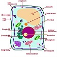
What is a cell, history of cell biology; prokaryotic and eukaryotic cells; cell shape and size; cell structure; the nucleus; the nucleolus; euchromatin and heterochromatin; the animal cell; the plant cell; human cells.
-
Cell Chemistry
-
Cell chemical composition; carbohydrates; lipids; nucleic acids; proteins; enzymes; cell membranes; golgi apparatus.
-
DNA, Chromosomes and Genes
-
DNA, Chromosomes, Genes; DNA replication; telomeres and telomerase; genetics; case study in genetic inheritance; phenotype and genotype; gene mutations.
-
Cell Division: Meiosis and Mitosis
-
Mitosis and meiosis overview; mitosis; meiosis.
-
Cell Membranes
-
Membranes; structure of cell membranes; movement of molecules through cell membranes; endocytosis; osmosis and filtration; hydrostatic pressure; active transport; electro-chemical gradient; nutrient and waste exhange in animal cells; mediated and non-mediated transport.
-
Protein Structure and Function
-
Protein structure; fibrous proteins; globular proteins; protein organisation; primary to quaternary structure; protein function.
-
Protein Synthesis
-
Overview; the function of ribonucleic acid in protein synthesis; transcription and translation; initiation; elongation; termination.
-
Food, Energy, Catalysis and Biosynthesis
-
Sources of energy; metabolism within the cell; catabolic metabolism; anabolic metabolism; ATP movement; Kreb's cycle; production and storage of energy; energy production pathways from different foods; biosynthesis of cell molecules; mitochondria; chloroplasts.
-
Intracellular Compartments, Transport and Cell Communication
-
Cell communication; endocrine signalling; paracrine signalling; autocrine signalling; cytoskeleton; actin filaments; intermediate filaments; microtubules.
-
Cell Cycle and Tissue Formation
-
The cell cycle; phases of the cell cycle; cell cycle regulation; cell death; cells to bodies; stem cells; animal tissues including muscle, connective, epithelial, nerve; blood.
Aims
-
Review basic cell structure and discuss the scope and nature of cell biology.
-
Describe the chemical components and processes of cells.
-
Describe the storage of genetic information within cells and how this information is passed on to the next generation.
-
Describe key concepts in molecular biology.
-
Discuss membrane structure and transport across cell membranes.
-
Discuss protein structure and function.
-
Describe and discuss protein synthesis.
-
Describe the significant processes involved in transfer and storage of energy in a cell.
-
Describe the significant processes that occur in cell communication and intracellular transport
-
Describe the life cycle of cells and how they combine to create different types of tissues.
How Do We Study Cells?
While there are a variety of ways to study cells; a lot of what we know and observe comes from using microscopes.
Microscopes allow us to observe things that are too small for the unaided human eye to see.
There are three types of microscopes:
- Light microscopes - bounce light through air or oil, onto and off the specimen.
- Electron microscopes - bounce a stream of electrons onto and off the specimen through a vacuum chamber.
- Helium ion microscopes (HIMs) - use focused beams of helium ions rather than electrons for surface imaging and analysis. The advantage of this is that because helium ions can be focused into a much smaller probe size (without burning or damaging the study specimen), it has improved focus depth and material contrast compared to electron microscopes, and better enables the user to determine the location of an edge of a critical feature.
Light and electron microscopes are older technology than helium. They both give very useful images for microbiology; but both can also cause damage to the specimen as they examine it. Helium microscopes have the advantage of not damaging the specimen in the same way.
Light Microscopes
 The first light microscopes were simply lenses that bent light as it bounced of what you were observing, and into your eye. This may work to some degree if you are in a well-lit place and the magnification was not great; but in order to see the smallest of things, you need greater magnification and immensely more intense light bouncing off what you are looking at. Hence a strong artificial light source becomes necessary. This type of microscope is relatively inexpensive but limited in two ways:
- Intense light can damage, hence may distort what you are looking at
- Lenses become more expensive to create and difficult to achieve a good focus as the magnification gets high
Oil Immersion Lens
This allows light to pass from a glass lens through oil; and in doing so there is less refraction of the stream of light. This enables a wider beam of light to be used thus increasing: numerical aperture, image brightness and resolving power. This in effect allows you to see smaller things (magnification up to x1450) with a better clarity.
TYPES OF LIGHT MICROSCOPES
There are three types of microscopes that allow you to see small things by bouncing light off those subjects.
- Stereoscope (or Stereo Microscope)
- Compound Microscope
- Confocal Microscope
Stereoscope (or Stereo Microscope)
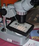
A stereo microscope is widely used in science. It allows for a magnified image to be viewed and is predominantly used for studying larger specimens as there is a limitation to the strength of magnification. For example, it is ideal for studying insects, plant material, some fungal samples and soil samples. Its use in microbiological applications is extremely limited as it doesn’t off resolution or magnification to adequately view micro-organisms.
Compound Microscope
A compound microscope includes a number of lenses and is a commonly used microbiology laboratories. The light enters from the base of the microscope through the condenser, through the specimen on a glass slide into the objective lens. The objective and ocular lens magnify the image before it can be seen through the eye piece. The light passing through the specimen can be controlled by the iris diaphragm. When using objective lens of higher magnification, more light is required to see the image clearly.
Dark-Field Microscopy is used to examine organisms that are light-sensitive and are better seen with a darker background. This microscope contains a condenser that prevents light being transmitted directly through the specimen, but causes the light to be reflected at an angle. The image seen is of a light specimen on a dark background.
Phase-Contrast Microscopy is generally required to observe micro-organisms live and unstained. Stains that are generally used to observe micro-organisms cause cell-death, and therefore they cannot be used on live samples. A phase-contrast microscope is fitted with a specialised condenser and objective lens so that the slightest changes in refracted light can be used to observe live specimens.
Fluorescence Microscopy is routinely used in diagnostic laboratories for identification purposes, whereby the fluorescent molecules are excited using ultraviolet light. Fluorescent dye molecules are attached (or tagged) to Antibody molecules, and will combine with Antigens if present in a specimen. Multiplexing of a number of fluorescence dyes can be used in these methods.
Confocal Microscope
Confocal microscopy is carried out through the use of ultraviolet laser beams that can be targeted through very small holes or slits. The fluorescence is then tightly focused, with the imagine being reconstructed by a computer to provide a high resolution image. A confocal microscope can pass through a thicker specimen at successive planes, providing three-dimensional images.
WHY STUDY THIS COURSE?
This course may be used to fill in gaps in your knowledge, or it may be the foundation for further learning.
Graduates from this course will have a foundation for learning more and working in any of the life sciences; with plants, animals, humans or any other types of organisms. Some will use this courses as the starting point for further formal studies., Others may go no further with their formal education, but nevertheless may continue to build on what they learn here.
Who might consider this course:
- Anyone working or interacting with biological scientists
- Technicians or lab assistants
- Anyone working in health, veterinary or agricultural industries
- Anyone with a desire to understand the microscopic world which impacts so much on our daily lives.
